

What Is Dynamic Image Analysis (DIA) and How Does It Compare to Micro-Flow Imaging?
Why image-based particle analysis is evolving beyond Micro-Flow Imaging into scalable DIA platforms
Particle characterization is a critical requirement across biopharmaceuticals, industrial fluids, fuels, and advanced materials. Over the past decade, image-based particle analysis has become increasingly important because it provides more information than legacy methods like light obscuration (LO), laser diffraction, or dynamic light scattering. Among these image-based technologies, two approaches are frequently discussed:
-
Micro-flow imaging (MFI®) — widely used in biopharmaceutical research
-
Dynamic Image Analysis (DIA) — a broader and more scalable technology suitable for both research and high-throughput QC environments
Micro-Flow Imaging and Flow-Image Microscopy are commonly used by many equipment manufacturers. MFI® happens to be a registered trademark of ProteinSimple (a Bio-Techne brand).
Although both methods, Micro-Flow Imaging and Flow-Image Microscopy, capture particle images in flow, their design philosophy, workflow suitability, and scalability are fundamentally different from that of Dynamic Image Analysis.
This post explains those differences — and why DIA is becoming the preferred solution when labs need sub-visible particle data that is accurate, scalable, and production-ready.
Micro-Flow Imaging: A Research-Focused Tool Built Around Very Low Sample Volumes
Micro-flow imaging was developed to help researchers study protein aggregation and other sub-visible particles in formulation development. Because early-stage biopharma work often involves small batch sizes, MFI systems were optimized for:
-
Microliter-scale sample consumption
- Low particle loads
This makes MFI highly effective for answering research questions such as:
-
“How does this formulation behave under freeze–thaw stress?”
-
“Are these protein aggregates reversible or irreversible?”
-
“Do silicone oil droplets appear during agitation?”
However, these design choices also introduce workflow limitations for scale-up efforts such as Quality Control, Manufacturing, and high-volume testing.
Real-World Research, QC, and Manufacturing Environments
1. Microliter sample volumes are not compatible with real-world sampling
Quality Control and release testing often require representative sample volumes, not tiny microliter aliquots. Low sample volumes and slow flow rates can limit:
-
Scalable sampling from IV bags
-
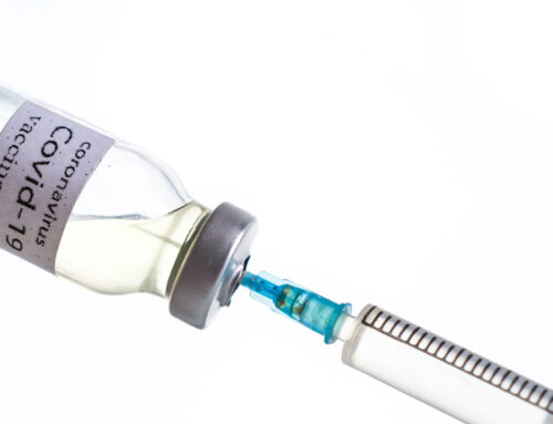
Testing from prefilled syringes
-
Homogeneous sampling of larger containers
-
High-volume batch screening in manufacturing
2. Low-throughput design
Low flow rates make it:
-
Slow for large sample sets
-
Difficult to integrate into release testing workflows
-
Inefficient for continuous or in-process monitoring
3. Sensitivity to particle load
Real biopharma samples often contain:
-
Surfactant micelles
-
Silicone droplets
-
Excipient crystals
-
Aggregates
-
Air bubbles
Low sample volume and slow flow rates will result in samples settling out, or rising to the top of the sample entry port and escaping analysis.
4. Slow flow rates allow particles to settle before imaging
Micro-flow imaging systems typically operate at very low flow rates to maintain image sharpness within the small optical cell. However, slow flow movement creates a physical problem:
-
Particles with even modest density differences (protein aggregates, silicone droplets, glass shards, polymer fragments) begin to settle or rise in the syringe or inlet tubing before they ever reach the imaging window.
-
Surface-active particles such as protein aggregates and silicone droplets may adhere to tubing walls, never entering the flow cell.
This means the population that reaches the camera may not match the population that is actually in the container, compromising representativeness.
6. Low sample volumes cannot produce statistically representative Sub-visible Particle data
In real-world biopharmaceutical sampling, particle distributions are often heterogeneous, especially in:
-
Prefilled syringes
-
IV bags
-
Bulk drug substance containers
-
Final drug product batches
Using only microliters to a few milliliters — typical for micro-flow imaging workflows — dramatically reduces the chance that the sample drawn accurately represents the full container.
Small volumes increase the likelihood of:
-
Missing rare but critical particle types
-
Underestimating or overestimating true sub-visible particle counts
-
Sampling error from stratification or settling
-
Failing to capture intermittent or low-frequency contaminants
-
Missing glass particulates from stopper coring or needle abrasion
For Quality Control and release testing, representative sampling is critical — and this is a key limitation of micro-flow imaging.
Dynamic Image Analysis (DIA): A Scalable, Production-Ready Alternative
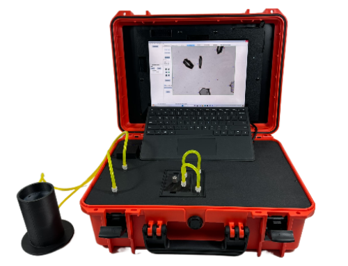
Dynamic Image Analysis solves the scalability and workflow limitations inherent in micro-flow imaging. Although both technologies image particles while in motion, DIA was developed to handle a much broader range of sample types, volumes, viscosities, particle concentrations, and workflows.
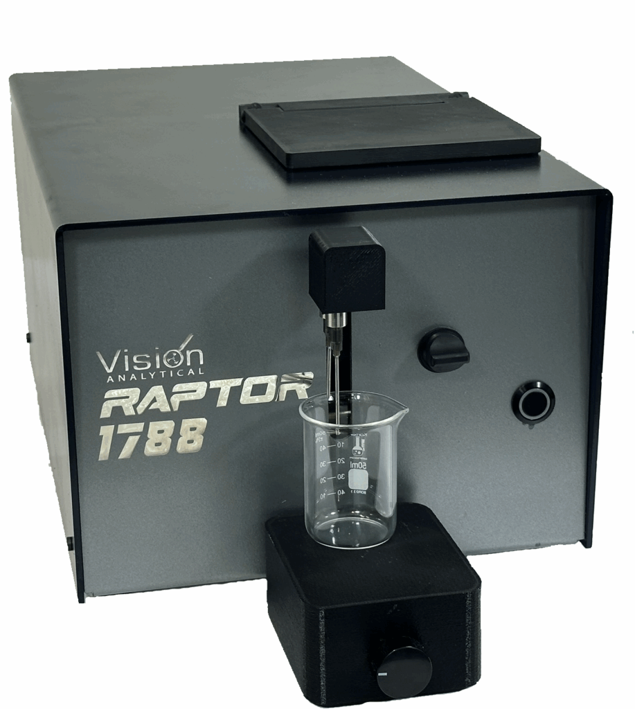
Systems like the Raptor 1788 bring the following advantages:
1. Works with Both Microliter and High-Volume Samples
DIA is inherently flexible. You can:
-
Analyze microliters when needed (similar to Micro-Flow Imaging), but also
-
Run milliliters to hundreds of milliliters for highly representative sampling
This allows direct measurement of:
-
Prefilled syringes (non-destructive testing)
-
IV bags (non-destructive testing)
-
Bulk drug substance containers
-
In-process streams
-
Larger manufacturing batches
This is one of the most decisive advantages over Micro-Flow Imaging and Flow-Image Microscopy.
2. Supports True “Real-World” Sampling — Including In-Process
Unlike Micro-Flow Imaging, which requires the sample to be brought to the instrument in very small quantities, DIA systems are designed to integrate with:
-
Light Obscuration particle counter sampling loops
-
In-process monitoring ports
-
Continuous flow systems
-
Syringe pump interfaces
- Small volume syringes
-
Larger industrial reservoirs
This offers major workflow benefits in QC and manufacturing:
-
Fewer sampling artifacts
-
Better representativeness
-
Fewer transfers, reducing contamination risk
-
Compatibility with automated sampling systems
3. Higher Throughput Without Sacrificing Image Quality
Modern DIA systems maintain strong image quality but allow:
-
Larger volumes
-
Higher particle concentrations
-
Faster flow rates
-
Continuous or semi-continuous sampling
This makes DIA suitable for:
-
Batch release testing
-
Stability testing
-
Line clearance verification
-
Routine contamination monitoring
4. Broad and Accurate Morphology Characterization
Both micro-flow imaging and DIA provide morphology — but DIA typically provides a more comprehensive feature set:
-
Aspect ratio
-
Circularity
-
Solidity
-
Convexity
-
Fiber length/width
-
Particle type classification (typically based on 30+ shape models)
- Particle Correlation Plots to find rare events
The diversity of shape metrics helps distinguish:
-
Aggregates vs silicone droplets
-
Glass shards vs polymer fragments
-
Fibers vs elongated protein clusters
-
Bubbles vs true particles
For root-cause analysis, this is often the deciding factor.
Why DIA Is Emerging as the Stronger Solution for Sub-Visible Applications
✔ Better sample representativeness
Because DIA can run larger volumes, it produces more statistically meaningful Sub-Visible Particle data.
✔ Suitable for QC, release testing, and manufacturing
Where Micro-Flow Imaging can be too slow, DIA adapts and scales.
✔ Handles real-world matrices
Surfactants, high particle loads, or viscous formulations do not stop DIA.
✔ Eliminates workflow bottlenecks
Fewer dilutions, fewer clogs, far less downtime.
✔ Enables direct sampling from production equipment
✔ Better accuracy through higher flow rates and anti-settling motion
DIA systems operate at significantly higher flow rates, preventing the sedimentation or flotation effects that occur in slow-flow systems.
This ensures that:
-
A broader spectrum of particles reaches the imaging zone
-
Fragile aggregates are transported consistently
-
Wall adhesion artifacts are reduced
-
The sample remains well-suspended and representative
The resulting data sets are more accurate and more reflective of the true particle load.
✔ Representative sample volumes reduce statistical error
Because DIA can process milliliters to hundreds of milliliters when needed, the sample measured is far more representative of the actual batch or container, drastically reducing sampling error — a critical factor in sub-visible particle decision making.
A major advantage for biologics manufacturing, where continuous inspection and in-line QC are increasingly valued.
Conclusion: Micro flow imaging Is Valuable, But DIA Is the Future
Micro-flow imaging remains a powerful research tool for studying protein aggregation. However, its low-volume, low-throughput design makes it challenging for high-volume or production environments.
Dynamic Image Analysis provides all the advantages of micro flow imaging while removing its limitations.
DIA allows laboratories to:
-
Test real-world sample volumes
-
Sample directly from production systems
-
Scale from R&D to QC to manufacturing
-
Handle challenging particle loads
-
Gain richer morphological insight
- Connect to Light Obscuration systems as an orthogonal method.
For organizations seeking robust, scalable, and representative sub-visible particle analysis, DIA is increasingly becoming the preferred — and more practical — option.
Trademark Attribution
MFI® is a registered trademark of ProteinSimple (a Bio-Techne brand). Any use in this article is for descriptive and comparative purposes only.
Learn More or Book a Demo
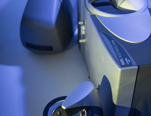
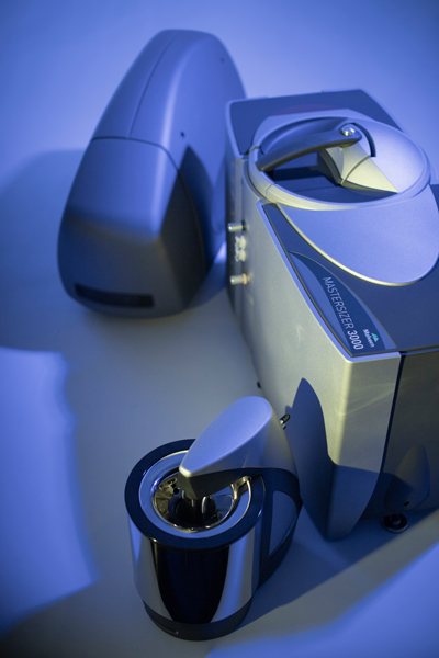
Visit www.ParticleShape.com to learn how Vision Analytical’s Dynamic Image Analysis solutions can enhance your existing Mastersizer system.

