
Application Note | Vision Analytical Inc. | Revised November 2026
 ]
Purpose
Stem cells are undifferentiated cells that can develop into specialized cell types, which makes characterizing them a critical part of therapeutic research and cell therapy development. This application note describes how Dynamic Image Analysis with the Raptor 1788 measures stem cell size, shape, opacity, and concentration in a single automated analysis, giving researchers quantitative data to support differentiation studies, population classification, and process development.
Why Morphology Matters in Stem Cell Characterization
As stem cells progress toward differentiation or apoptosis, their physical properties change. Cell morphology, including size, circularity, and opacity, can serve as an indicator of cell state. Traditional particle sizing techniques report only size and assume every particle is a sphere, which limits their usefulness for biological cell populations that contain subpopulations of varying shape and darkness. Manual microscopy shows what the cells look like, but it is low-throughput and depends on the operator.
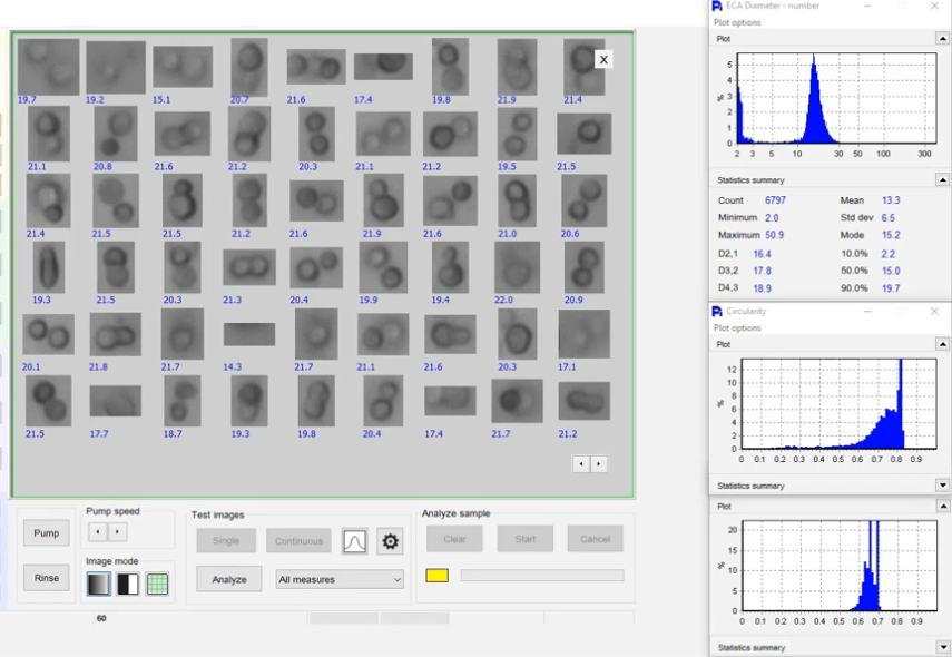
Automated Dynamic Image Analysis addresses both limitations. As cells pass through the instrument’s detection zone, images are captured and analyzed in real time, generating size and shape distributions across thousands of cells per minute. That throughput, combined with a thumbnail image of every cell for visual confirmation, makes the technique well suited to stem cell characterization in research and process development settings.
Instrument Overview: Raptor 1788 Dynamic Image Analyzer
The Raptor 1788 is a dynamic image analyzer that measures particle size and shape simultaneously. For stem cell work, the instrument captures a thumbnail of every measured cell and organizes the thumbnails for review after the analysis. Data can be exported directly to Excel for further statistical analysis, and overlays can be generated to compare populations across samples or time points.
Measuring Stem Cell Size, Circularity, Opacity and Concentration
Size
The cell diameter distribution across the measured population, reported as equivalent circular diameter (ECD).
Circularity
A measure of how round each cell is. Cells that become more elongated, as may occur during differentiation or progression toward apoptosis, show a decrease in circularity over time, which makes circularity a useful morphological indicator of cell state.
Opacity
A measure of how dark each cell appears in the image. Opacity differences can reveal subpopulations within a sample, such as live versus dead cells or cells at different stages of differentiation. The Raptor 1788 is not a dedicated viability analyzer, but opacity distributions provide a useful first-pass indication of population heterogeneity.
Concentration
The instrument reports particles per milliliter, giving a count of the cell population. Cluster rejection settings can be adjusted to minimize the contribution of cell aggregates to the concentration result.
Correlation Plots, Rare Event Detection and Cell Classification
Correlation Plots
Any two measured parameters, such as size and opacity, can be plotted against each other to test hypotheses about cell population behavior. For example, a researcher can ask whether cell size correlates with darkness across a population, and identify outliers that may represent a biologically distinct subpopulation.
Rare Event Detection
Because the Raptor 1788 captures a thumbnail image of every measured particle, individual cells of interest can be found within a population of 50,000 or more. A cell with unusually high circularity, extreme size, or anomalous opacity, which would be invisible in a bulk histogram, can be located and examined directly. This would be impractical with bulk measurement techniques or manual image review.
Particle Classification
The classification feature segments the population into user-defined classes based on any combination of size and shape parameters. Example classes for stem cell work might include:
- Small round cells
- Large round cells
- Elongated cells
- Dark cells
- Light cells
- Unclassified
Each class is reported with particle count, percentage of the total population, mean size, and standard deviation, giving a full statistical breakdown of each morphological subpopulation on its own.
Video: Stem Cell Analysis on the Raptor 1788
This demonstration shows the Raptor 1788 measuring a stem cell sample, from live image capture to thumbnail review and size and shape results.
Conclusion
Dynamic Image Analysis with the Raptor 1788 characterizes stem cell populations in a single automated measurement. Thumbnail review gives visual confirmation of cell morphology, while correlation plots, particle classification, rare event detection, and Excel export provide the quantitative data needed for research documentation and process development. Combining size, shape, opacity, and concentration in one analysis makes the Raptor 1788 a practical tool for researchers working with stem cells and other biological cell populations.
Frequently Asked Questions
How is Dynamic Image Analysis different from a standard cell counter?
Most cell counters report count and size and assume every cell is a sphere. Dynamic Image Analysis captures an image of every cell, so in addition to size and concentration it measures shape (circularity, elongation) and opacity, and lets you see each cell to confirm what was counted.
Can Dynamic Image Analysis distinguish live and dead stem cells?
Opacity differences can reveal subpopulations such as live and dead cells, and they give a useful first-pass view of population heterogeneity. The Raptor 1788 is not a dedicated viability analyzer, so where a formal viability result is required it should be confirmed with an established viability method.
How many cells are measured in a sample?
The Raptor 1788 analyzes thousands of cells per minute, and populations of 50,000 cells or more can be measured and reviewed individually, which is what makes rare event detection possible.
Can stem cell morphology be tracked over time?
Yes. Results from different samples or time points can be overlaid, so changes in size, circularity, or opacity during differentiation or culture can be compared directly. All data can also be exported to Excel for further analysis.
Related Resources
- Particle Shape and Size Analysis for Stem Cell Research (blog overview)
- How particle shape and size analysis supports stem cell research (white paper on News-Medical)
- How Dynamic Image Analysis Works
- USP <788> and <1788> Compliance Guide (for pharmaceutical particle applications)
Test Your Stem Cell Samples
Want to see what Dynamic Image Analysis reveals about your cells? Send us a sample and we’ll run it on the Raptor 1788 and share the thumbnails, size and shape distributions, and classification results.